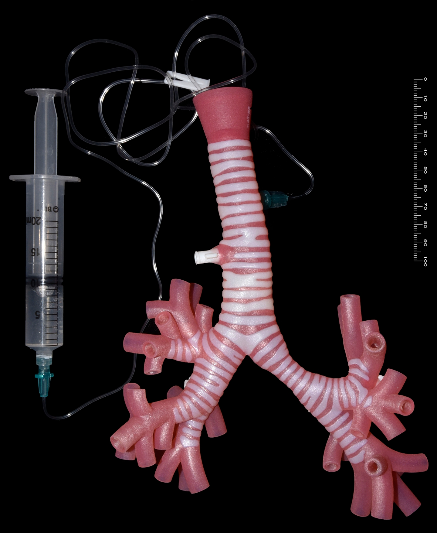
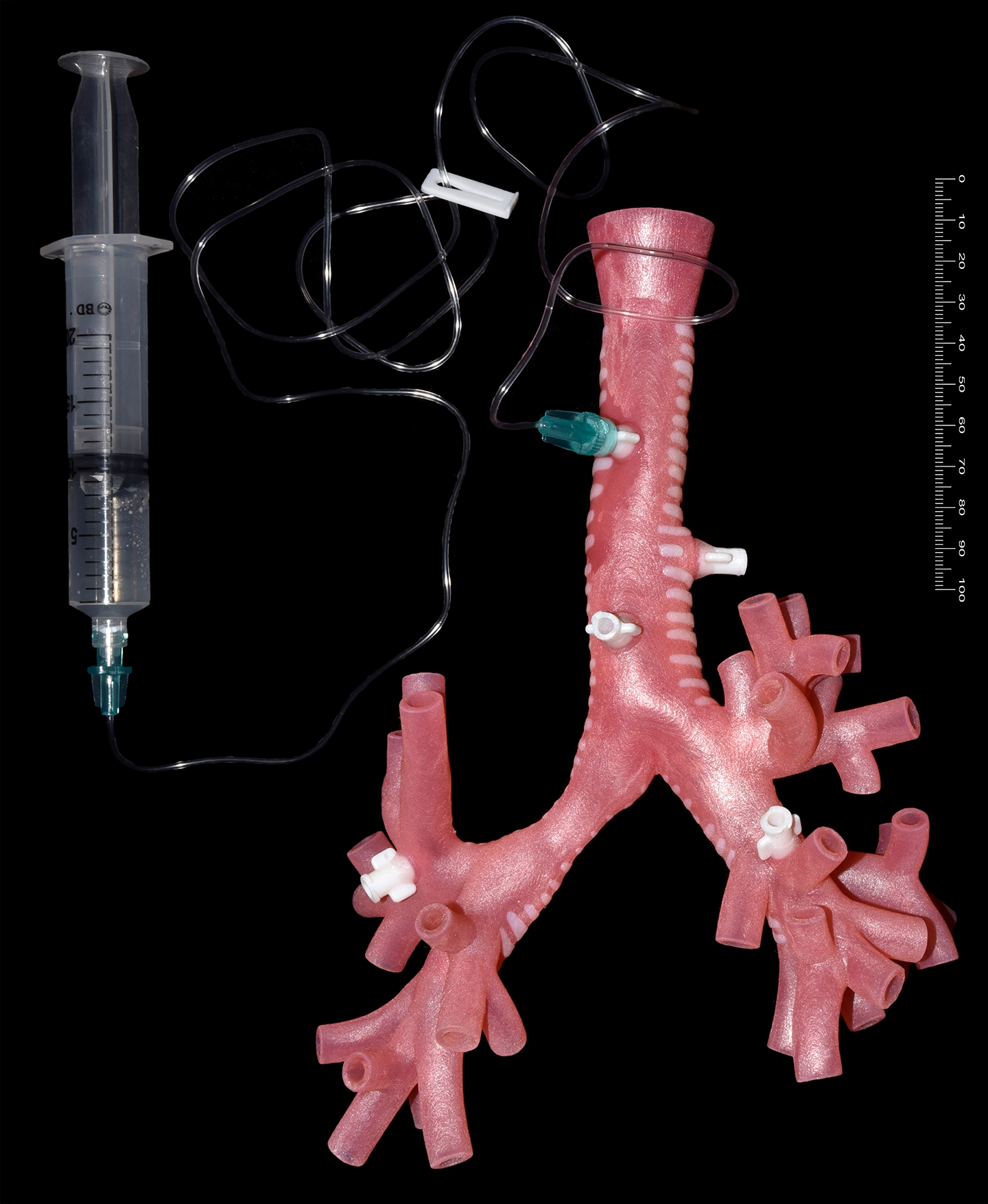
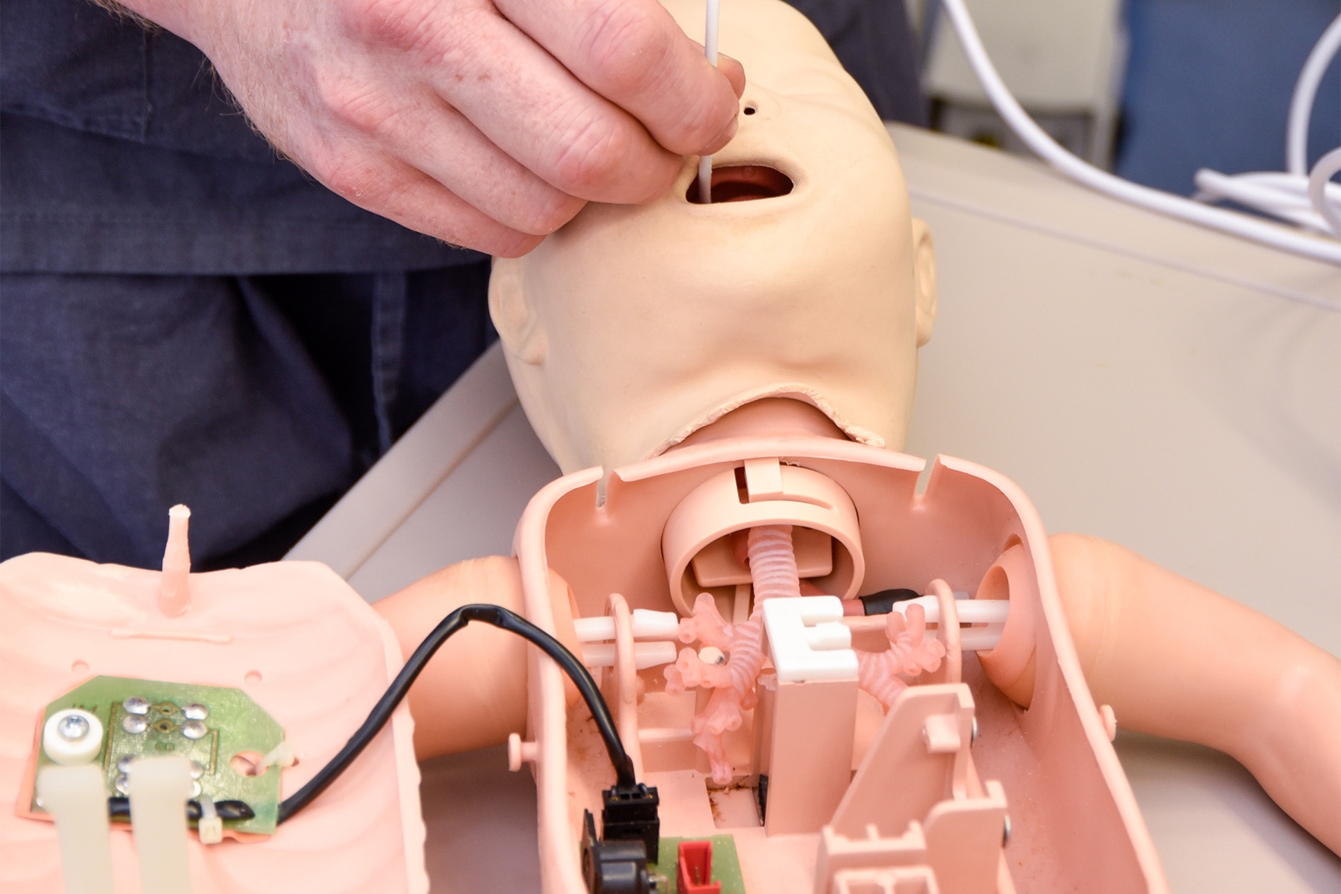
Interactive models
Moving beyond simple anatomy, ethical and future-focussed design goals inspired us to develop responsive models. Our models are used by simulation centres for advanced skill development, operative awareness and clinician response. Companies also use our models to facilitate R&D testing and evaluation of new product concepts. Our 3D-printed adult trachea is currently exhibited at the Museum of Anaesthesia.
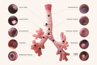
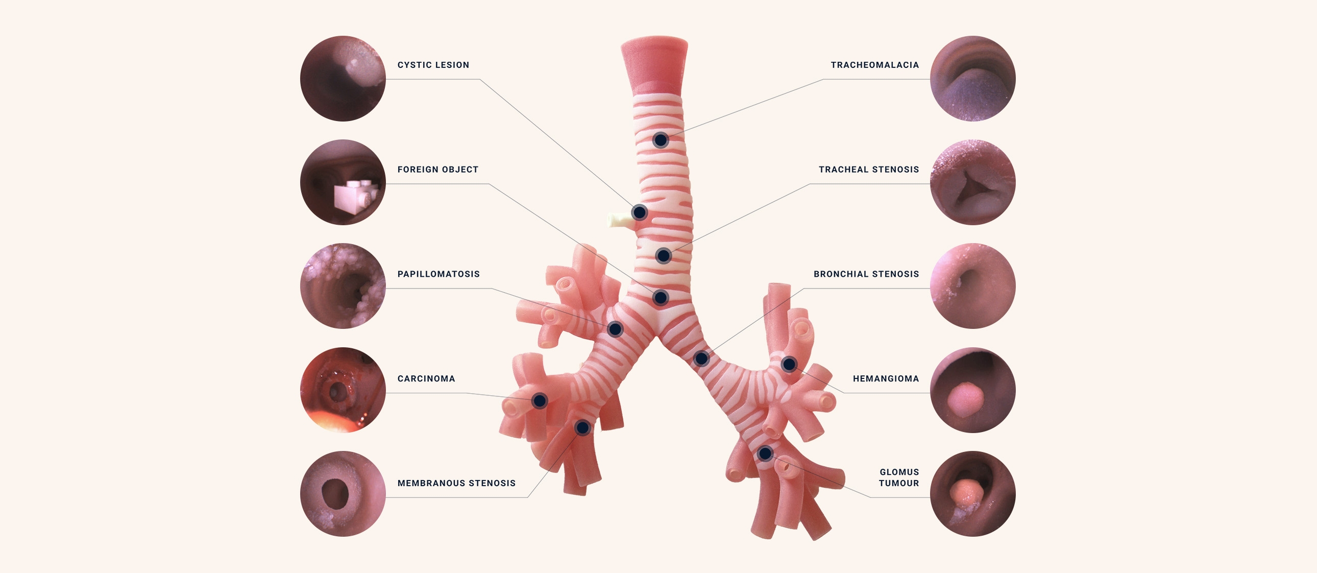
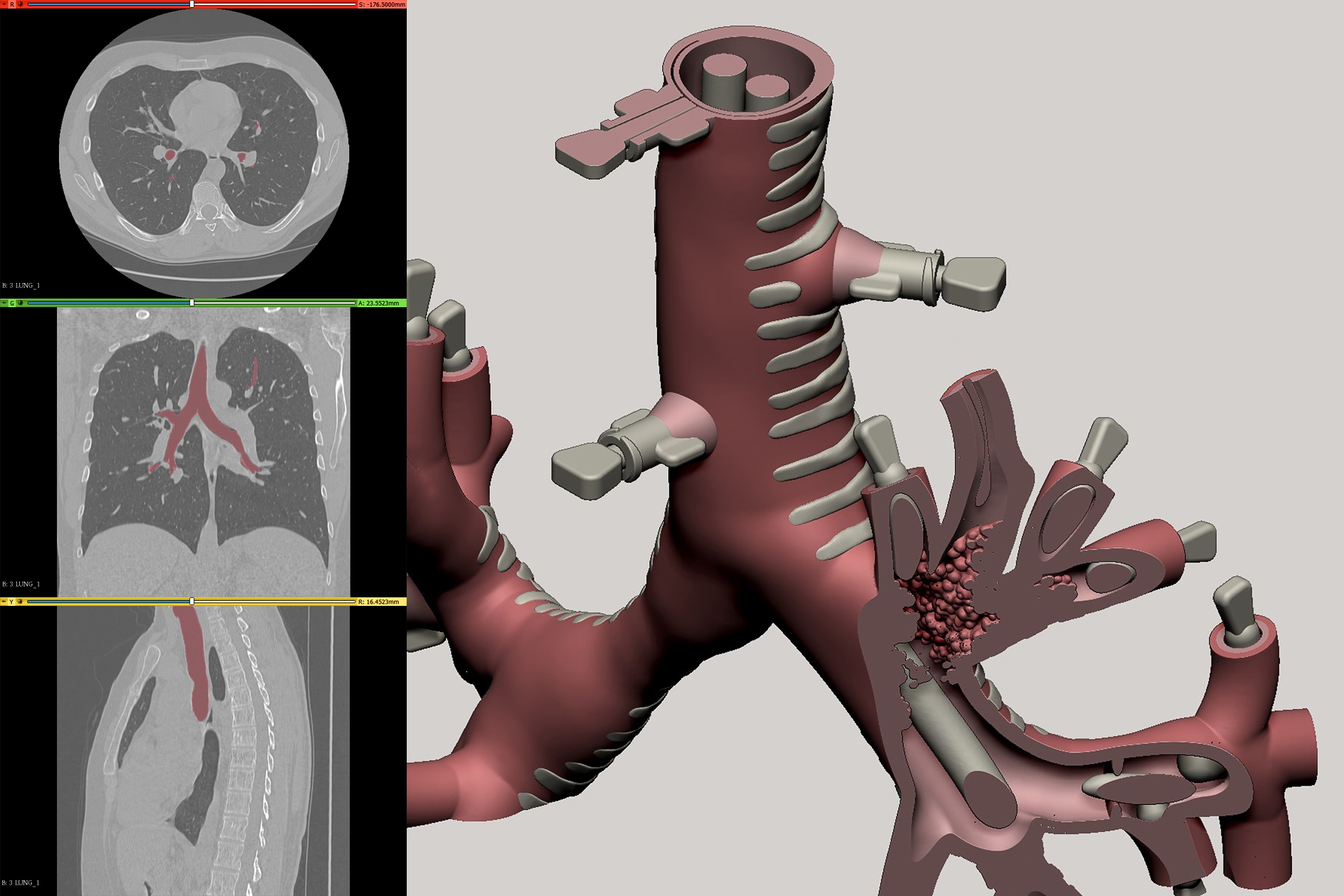
Patient pathology
We work with Dicom images and observation to facilitate the best equivalence of shape, colour, activity and realism.
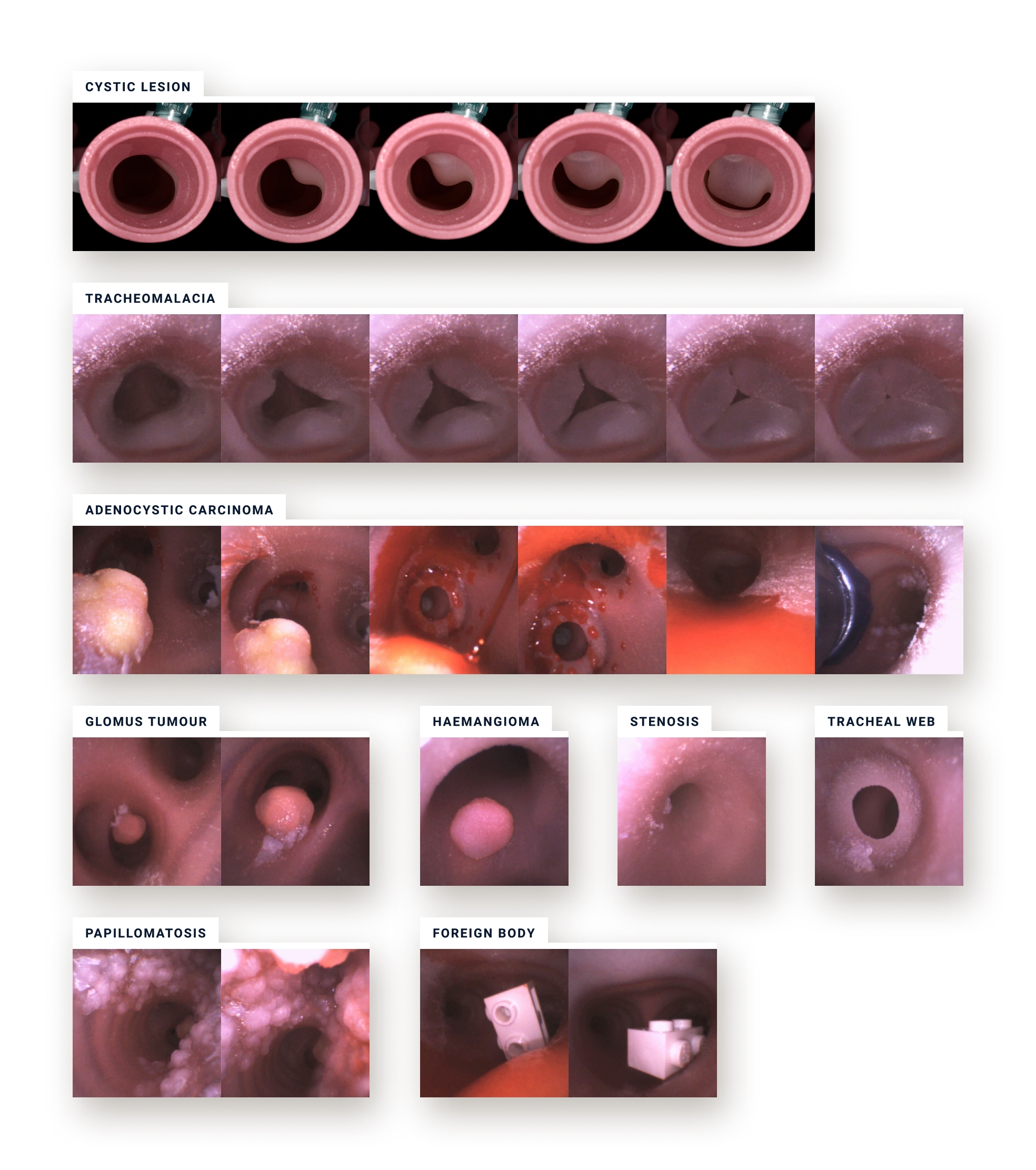
Through the scope
Pathology can be placed where required within the model. Levels of complexity, interaction and dynamics can also be tailored to requirements.
The images below show one of our trachea models in use during a simulated bronchoscopy.
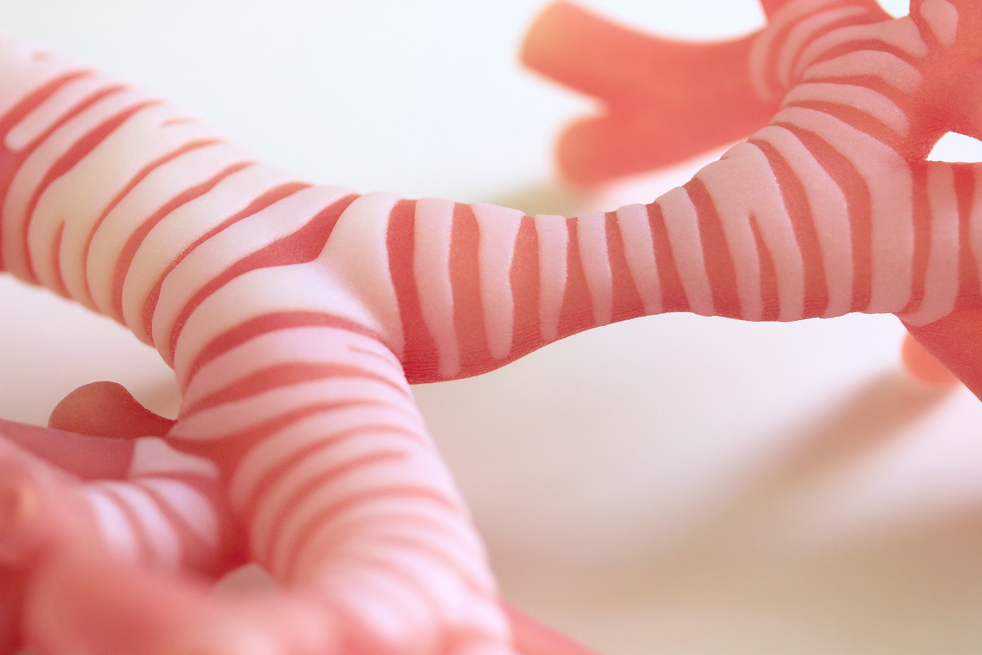
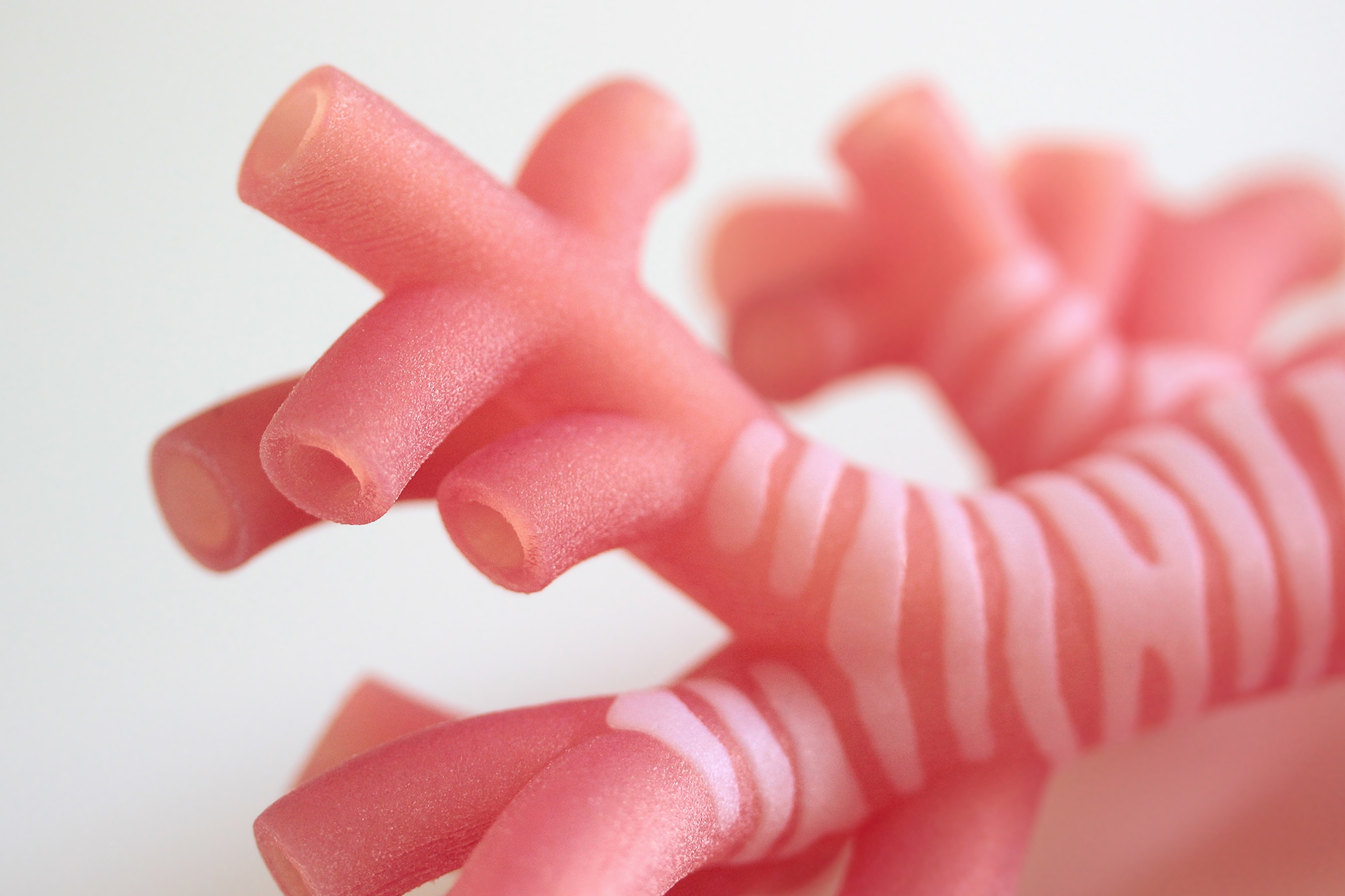
Material behaviour and longevity
Our products are 3D printed in a multi-property polymer. In effect, we vary the density and softness of regions to allow a true likeness to anatomy, from adult to infant. Careful handling and care will allow models to function well for extended periods.